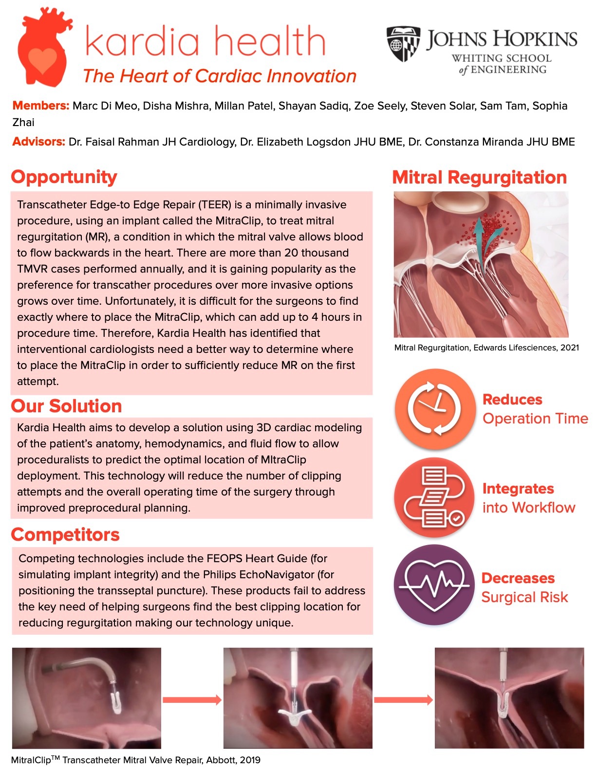
A better placement of the MitraClip to the Mitral Valve leaflets during a Transcatheter Edge to Edge Repair (TEER)
Team: BME Design Team: Kardia Health
- Program: Biomedical Engineering
- Course:
Project Description:
The mitral valve sits within the heart and consists of two leaflets that open and close during the cardiac cycle. It acts to prevent the reversal of blood flow from the left ventricle to the left atrium. Failure of the mitral valve to close tightly results in mitral regurgitation, which can lead to heart failure and mortality. One treatment of severe mitral regurgitation is transcatheter edge-to-edge repair (TEER) with the MitraClip NTR/XTR Clip Delivery System by Abbott. This is a minimally invasive, catheter-based procedure typically performed on high-risk patients with confounding health conditions. The MitraClip device grasps the mitral valve leaflets that are allowing the backflow of blood. Accurate clip positioning and deployment requires continuous evaluation of 2D and 3D TEE, fluoroscopy and hemodynamic data. The decision to place a clip in a certain location is based on multimodality imaging and clinician experience. However, the impact of the clip is unknown until the clip is fully deployed, especially when the images referenced intraoperatively are not clear or in the desired view. Furthermore, clinicians report that misplacement of the MitraClip can result in valve torsion, single leaflet attachment, and clip engagement with subvalvular structures, which can cause significant mitral valve obstruction. This can lead to the need for reclipping several times and/or multiple clips, increasing the procedure time by an estimated 20-30 minutes per reclipping. Considering that the majority of TEER patients are at high risk for morbidity and mortality, reducing procedure time and increasing accuracy of MitraClip placement by any amount is crucial. In order to better guide the MitraClip, our team is creating an image analysis software that can display an indicator of where MitraClip placement should occur across images from different modalities. Prior to the procedure, surgeons will consult 2D and 3D images of the heart and mark the location where they wish to place a MitraClip. Our algorithm will then place an indicator at that location and will display the indicator intra-operatively, marking the target location for the MitraClip on the live 2D and 3D TEE images. This indicator will update in real-time to enable surgeons to keep track of the desired location for clip placement as the imaging modalities and views of the mitral valve change.


