Eliminating Repeat Procedures to Treat Biliary Strictures
Team: BiliFlow
- Program: Biomedical Engineering
- Course: BME Undergraduate Design Team
Project Description:
A biliary stricture is an abnormal narrowing of the bile duct which impedes bile flow, usually caused by a surgical error or malignancy in surrounding organs. According to the National Inpatient Sample, there were approximately 101,000 biliary dilation procedures performed in 2019. The current treatment is to repeatedly upsize drainage catheters through the biliary stricture, up to five times every two weeks, to restore bile flow and fully dilate the stricture. Repeat procedures are performed to prevent liver damage: to reach the bile duct, the drainage catheter must pass through the liver, which is exposed to blunt trauma when a drainage catheter that is too large is inserted through it; however, if the drainage catheters are gradually upsized, there is time for the liver to adjust to the drainage catheter size. Unfortunately, these repeat procedures mean more pain and hospital time for patients, more labor for interventional radiologists, and lost profit for hospitals and insurance companies. To address this, we developed a novel device, the BiliFlow Sheath: a device that performs stricture dilation in a single procedure, features drainage and securement mechanisms for long-term deployment, adapts to different biliary morphologies, and permits balloon targeting to stricture locations. With bench-top testing, we demonstrated that our device does not impede bile drainage, is adjustable to different stricture locations, and is easily maneuvered through a biliary tree model. The BiliFlow Sheath has the potential to change stricture treatment for the better by improving dilation procedure effectiveness and reducing the burden of repeat procedures.
Project Photo:
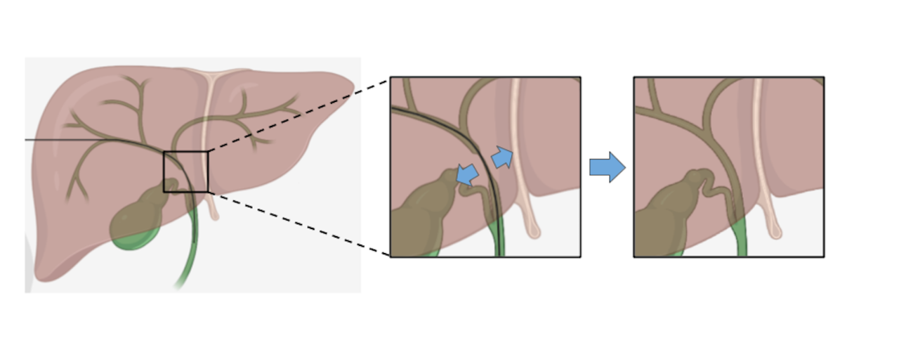
The left of the figure shows the liver and biliary system with a guidewire traveling through the biliary tree and passing a biliary stricture, which is a narrowing of the bile duct. In the middle is a zoomed in image showing the stricture at greater scale. Finally, at the right is a picture of a fully dilated stricture with normal morphology, which is what our solution will lead to.


