Optimizing skin cancer treatment with a rapid, slide-free margin assessment tool
Team: Advanced BME Design Team: OptiMohs
Program:
Biomedical Engineering
Project Description:
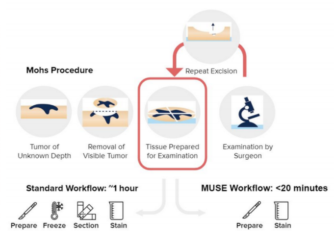
Mohs micrographic surgery (MMS) is the gold standard treatment for nonmelanoma skin cancer, one of the most common types of cancer. MMS has excellent patient outcomes, because the surgeon iteratively examines the entire surgical margin via histopathology, allowing the tumor to be excised completely. This process is repeated for each stage, leading to scheduling complications, high costs, a strain on hospital resources, and reduced accessibility for patients. Our goal is to acquire images quickly and at a low cost while maintaining the quality needed to inform iterative resections. While standard processing requires the tissue to be frozen and sliced onto slides, our solution will enable unsliced tissue to be imaged by integrating Microscopy with UV Surface Excitation (MUSE) into the Mohs workflow. By streamlining the most inefficient steps of the procedure, we will enable the gold standard of nonmelanoma skin cancer treatment to be accessible to more patients.
Team Members
-
[foreach 357]
-
[if 397 not_equal=””][/if 397][395]
[/foreach 357]
Project Mentors, Sponsors, and Partners
-
Project Mentor: Dr. Elise Ng, M.D.
-
Faculty Mentor: Dr. Nicholas Durr, Ph.D.
-
Dr. Ryan Niemeier, Ph.D.,
-
Dr. Greg Mckay, M.D., Ph.D.
Course Faculty
-
[foreach 429]
-
[if 433 not_equal=””][/if 433][431]
[/foreach 429]